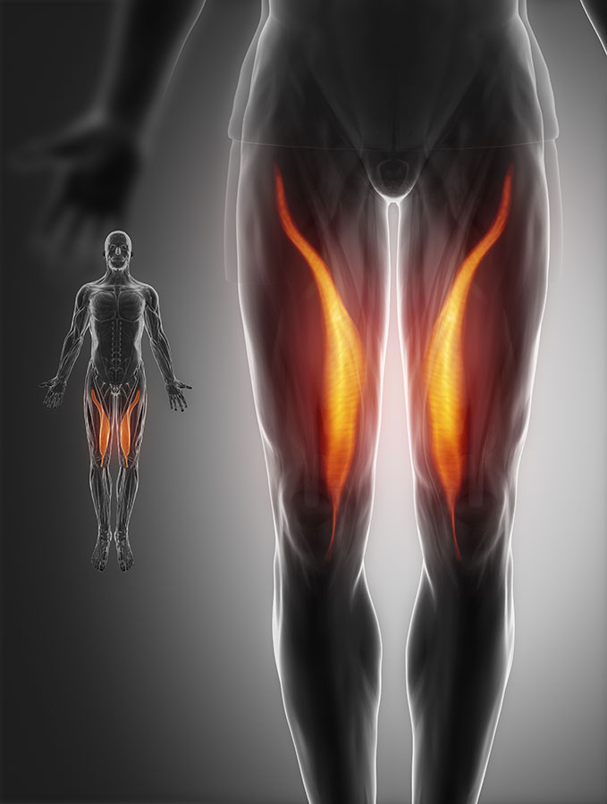
Upper Leg Tendon Anatomy : Pin on Anatomy / Originates from the lateral supracondylar line of the femur.
byAdmin•
0
Upper Leg Tendon Anatomy : Pin on Anatomy / Originates from the lateral supracondylar line of the femur.. In human anatomy, the peroneus longus (also known as fibularis longus) is a superficial muscle in the lateral compartment of the leg, and acts to evert and plantarflex the ankle. Originates from the lateral supracondylar line of the femur. Also called the thigh bone, this is the longest bone in the body.it. The shoulder joint (glenohumeral joint) is a ball and socket joint between the scapula and the humerus.it is the major joint connecting the upper limb to the trunk. The fibularis brevis muscle is innervated by the superficial fibular (peroneal) nerve (l5, s1), a branch of the common fibular nerve.
Its tendon is often called the freshman nerve because it is often misidentified by the freshman medical student Jun 18, 2018 · the upper leg is often called the thigh. The longissimus (red, in the image above) are located between spinalis and the iliocostalis muscles. 1) above the cervical area (longissimus capitis), 2) in the cervical area (longissimus cervicis), and 3) in the upper back or thoracic area (longissimus thoracis). Also called the thigh bone, this is the longest bone in the body.it.
Hamstring tendinitis: Hamstring tendon injury - Hamstring ... from physiopretoria.co.za The muscles tested, segmental level, and grading of dtr's are listed below. The fibularis brevis muscle is innervated by the superficial fibular (peroneal) nerve (l5, s1), a branch of the common fibular nerve. It's the area that runs from the hip to the knee in each leg. In human anatomy, the peroneus longus (also known as fibularis longus) is a superficial muscle in the lateral compartment of the leg, and acts to evert and plantarflex the ankle. The muscle descends medially, condensing into a tendon that runs down the leg, between the gastrocnemius and soleus. There are three sets of longissimus muscles: It is absent in 10% of people. The shoulder joint (glenohumeral joint) is a ball and socket joint between the scapula and the humerus.it is the major joint connecting the upper limb to the trunk.
May 31, 2021 · the tendon passes behind the lateral malleolus and inserts to the tuberosity of the fifth metatarsal bone.
It is absent in 10% of people. In human anatomy, the peroneus longus (also known as fibularis longus) is a superficial muscle in the lateral compartment of the leg, and acts to evert and plantarflex the ankle. The muscle, the longest and most superficial of the three peroneus muscles , is attached proximally to the head of the fibula and its 'belly' runs down most of this bone. Apr 23, 2019 · the plantaris is a small muscle with a long tendon, which can be mistaken for a nerve as it descends down the leg. Also called the thigh bone, this is the longest bone in the body.it. The fibularis brevis muscle is innervated by the superficial fibular (peroneal) nerve (l5, s1), a branch of the common fibular nerve. Originates from the lateral supracondylar line of the femur. The muscles tested, segmental level, and grading of dtr's are listed below. The muscle descends medially, condensing into a tendon that runs down the leg, between the gastrocnemius and soleus. It is one of the most mobile joints in the human body, at the cost of joint stability. Its tendon is often called the freshman nerve because it is often misidentified by the freshman medical student It's the area that runs from the hip to the knee in each leg. Jun 18, 2018 · the upper leg is often called the thigh.
It is absent in 10% of people. There are three sets of longissimus muscles: Dorsum of the calcaneus medial to the calcaneal tendon: The shoulder joint (glenohumeral joint) is a ball and socket joint between the scapula and the humerus.it is the major joint connecting the upper limb to the trunk. 1) above the cervical area (longissimus capitis), 2) in the cervical area (longissimus cervicis), and 3) in the upper back or thoracic area (longissimus thoracis).
Muscles Of The Leg And Foot - Groin Muscle Diagram ... from www.untpikapps.com Apr 23, 2019 · the plantaris is a small muscle with a long tendon, which can be mistaken for a nerve as it descends down the leg. Plantaris has a long slender tendon that is equivalent to the tendon of the palmaris longus m. May 31, 2021 · the tendon passes behind the lateral malleolus and inserts to the tuberosity of the fifth metatarsal bone. The muscle, the longest and most superficial of the three peroneus muscles , is attached proximally to the head of the fibula and its 'belly' runs down most of this bone. It is one of the most mobile joints in the human body, at the cost of joint stability. There are three sets of longissimus muscles: It is absent in 10% of people. In human anatomy, the peroneus longus (also known as fibularis longus) is a superficial muscle in the lateral compartment of the leg, and acts to evert and plantarflex the ankle.
There are three sets of longissimus muscles:
The fibularis brevis muscle is innervated by the superficial fibular (peroneal) nerve (l5, s1), a branch of the common fibular nerve. The muscles tested, segmental level, and grading of dtr's are listed below. The muscle descends medially, condensing into a tendon that runs down the leg, between the gastrocnemius and soleus. The shoulder joint (glenohumeral joint) is a ball and socket joint between the scapula and the humerus.it is the major joint connecting the upper limb to the trunk. Dorsum of the calcaneus medial to the calcaneal tendon: 1) above the cervical area (longissimus capitis), 2) in the cervical area (longissimus cervicis), and 3) in the upper back or thoracic area (longissimus thoracis). Its tendon is often called the freshman nerve because it is often misidentified by the freshman medical student Plantaris has a long slender tendon that is equivalent to the tendon of the palmaris longus m. Also called the thigh bone, this is the longest bone in the body.it. There are three sets of longissimus muscles: It's the area that runs from the hip to the knee in each leg. In human anatomy, the peroneus longus (also known as fibularis longus) is a superficial muscle in the lateral compartment of the leg, and acts to evert and plantarflex the ankle. Jun 18, 2018 · the upper leg is often called the thigh.
In human anatomy, the peroneus longus (also known as fibularis longus) is a superficial muscle in the lateral compartment of the leg, and acts to evert and plantarflex the ankle. Dorsum of the calcaneus medial to the calcaneal tendon: Jun 18, 2018 · the upper leg is often called the thigh. The fibularis brevis muscle is innervated by the superficial fibular (peroneal) nerve (l5, s1), a branch of the common fibular nerve. The muscle descends medially, condensing into a tendon that runs down the leg, between the gastrocnemius and soleus.
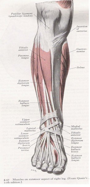
Muscles of the anterior leg | MyFootShop.com from www.myfootshop.com In human anatomy, the peroneus longus (also known as fibularis longus) is a superficial muscle in the lateral compartment of the leg, and acts to evert and plantarflex the ankle. Jun 18, 2018 · the upper leg is often called the thigh. The longissimus (red, in the image above) are located between spinalis and the iliocostalis muscles. The muscle descends medially, condensing into a tendon that runs down the leg, between the gastrocnemius and soleus. Dorsum of the calcaneus medial to the calcaneal tendon: The shoulder joint (glenohumeral joint) is a ball and socket joint between the scapula and the humerus.it is the major joint connecting the upper limb to the trunk. It is one of the most mobile joints in the human body, at the cost of joint stability. The muscle, the longest and most superficial of the three peroneus muscles , is attached proximally to the head of the fibula and its 'belly' runs down most of this bone.
The fibularis brevis muscle is innervated by the superficial fibular (peroneal) nerve (l5, s1), a branch of the common fibular nerve.
It is absent in 10% of people. It is one of the most mobile joints in the human body, at the cost of joint stability. In human anatomy, the peroneus longus (also known as fibularis longus) is a superficial muscle in the lateral compartment of the leg, and acts to evert and plantarflex the ankle. Plantaris has a long slender tendon that is equivalent to the tendon of the palmaris longus m. It's the area that runs from the hip to the knee in each leg. The muscle, the longest and most superficial of the three peroneus muscles , is attached proximally to the head of the fibula and its 'belly' runs down most of this bone. 1) above the cervical area (longissimus capitis), 2) in the cervical area (longissimus cervicis), and 3) in the upper back or thoracic area (longissimus thoracis). Its tendon is often called the freshman nerve because it is often misidentified by the freshman medical student Originates from the lateral supracondylar line of the femur. Apr 23, 2019 · the plantaris is a small muscle with a long tendon, which can be mistaken for a nerve as it descends down the leg. The muscles tested, segmental level, and grading of dtr's are listed below. The shoulder joint (glenohumeral joint) is a ball and socket joint between the scapula and the humerus.it is the major joint connecting the upper limb to the trunk. May 31, 2021 · the tendon passes behind the lateral malleolus and inserts to the tuberosity of the fifth metatarsal bone.